
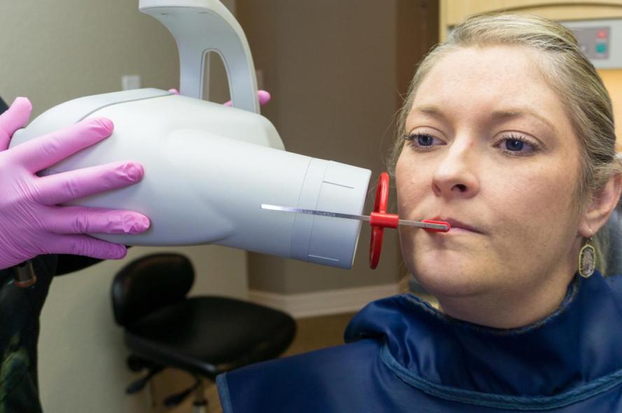
Dental X-rays sit high on the list of things patients worry about unnecessarily. The radiation involved is extremely low, but the questions keep coming up, especially from parents booking appointments for kids or patients who had a lot of imaging done years ago. Here is a clear, evidence-based answer.
Yes, Dental X-Rays Are Safe
Dental X-rays involve a genuinely small dose of radiation, and modern digital equipment has made that dose even lower than it was a decade ago. The concern most patients carry into the appointment is usually far larger than what the science actually supports.
According to Dr. Benavides of the American Dental Association, dental X-rays are safe, and in some cases a single dental X-ray delivers less radiation than a person receives in one day from natural background radiation simply by living in the world. That comparison alone reframes how small this exposure actually is.
The radiation dose from a dental X-ray is measured in microsieverts, generally falling between about 1 and 100 micro Sieverts depending on the type of X-ray taken, compared to roughly 10 microsieverts for a standard chest X-ray and around 5 microsieverts for a typical dental bitewing. In context, dental imaging sits well below many other routine medical scans.
Understanding these numbers is useful, but what matters more is how dental professionals have recently updated their approach to ordering X-rays in the first place.
New Guidelines Confirm X-Rays Should Be Used in Moderation
A Major Update From the ADA
In January 2026, the American Dental Association published its first new dental X-ray recommendations in more than a decade, confirming that X-rays should be ordered only when clinically necessary, in order to minimize radiation exposure for both patients and dental professionals.
These updated recommendations are also the first from the ADA to address both traditional 2D imaging and 3D cone beam computed tomography, or CBCT, together within a single framework covering specific clinical scenarios.
The Core Principle: Clinical Necessity, Not Routine Scheduling
At the heart of this update is a simple message: dental X-rays should be prescribed only when they are clinically justified, rather than relying on routine schedules or convenience.
Imaging decisions are meant to be based on a careful review of the patient’s medical and dental history, disease risk, prior images already on file, and current clinical findings.
This shift matters because it directly answers the most common patient question: why does my dentist sometimes skip X-rays at a visit where I expected them? The answer is that modern guidance actively discourages imaging that is not clinically justified. Let’s look at what this means for shielding practices as well.
Why Lead Aprons Are No Longer Standard
The Shielding Recommendation Has Changed
As of early 2024, the use of lead abdominal aprons or thyroid collars during dental X-rays is no longer recommended, according to an expert panel established by the American Dental Association’s Council on Scientific Affairs.
Thyroid collars and other shielding, such as lead aprons, can actually block the primary X-ray beam, which may result in the need for additional radiographs to capture the necessary diagnostic image.
The Science Behind the Change
This recommendation reflects evidence that modern dental imaging equipment already delivers such low radiation doses that additional shielding provides negligible protective benefit while sometimes interfering with image quality.
If you have noticed your dentist no longer offers a lead apron during your X-ray, this is why. It is not an oversight, it reflects updated, evidence-based safety guidance. Now let’s cover how digital technology has changed the safety picture even further.
How Digital X-Rays Have Reduced Radiation Exposure
Digital Sensors Require Less Radiation Than Film
Digital X-ray technology cuts radiation doses by up to 50 percent compared to older conventional film methods, making modern dental imaging significantly safer than what patients may remember from years past.
At Heritage Dental Studio, our digital X-ray equipment reflects this generational shift in imaging technology, offering clearer diagnostic images with meaningfully lower radiation exposure.
Additional Safety Measures Built Into Modern Practice
Collimating the X-ray beam to the specific region being examined, a technique called rectangular collimation, has been shown to reduce radiation dose consistently by more than 40 percent.
Additional measures, including the use of long position-indicating devices to maximize the distance between the radiation source and the patient’s skin, further reduce exposure during routine imaging.
These layered safety improvements mean that a modern dental X-ray delivers a fraction of the radiation exposure it would have decades ago. This naturally raises the question of how often you actually need this imaging.
How Often Do You Actually Need Dental X-Rays?
It Depends on Your Individual Risk Profile
The frequency of dental X-rays depends on individual oral health needs and risk factors, and patients are encouraged to talk directly with their dentist about their personal X-ray schedule.
There is no single universal answer, since a patient with a history of frequent cavities will need more frequent imaging than someone with excellent oral health and no restorative history.
General Patterns Dentists Follow
For most low-risk adult patients, bitewing X-rays are typically recommended every 12 to 24 months to check for decay between teeth that is not visible during a standard exam. Higher-risk patients, including those with a history of frequent cavities or gum disease, may need imaging more often.
Full Mouth Series and Panoramic Imaging
A full mouth series or panoramic X-ray is typically recommended less frequently, often every three to five years for patients with stable oral health, though this can vary depending on findings from your comprehensive exam.
New Patient Visits Often Require Baseline Imaging
If you are new to a clinic, your dentist will often want to establish a baseline set of images, since prior imaging from a previous provider may not always be available or current. This baseline helps track any changes over time during future visits.
Rather than following a rigid calendar, your dentist tailors imaging frequency to your specific clinical picture, which brings us to the specific scenarios where X-rays genuinely matter most.
When X-Rays Provide Real Diagnostic Value
Detecting Decay Between Teeth
Cavities that form between teeth are often invisible during a visual exam alone. X-rays are the most reliable way to catch this type of decay before it progresses significantly.
Evaluating Bone Health Around Teeth
X-rays reveal the condition of the bone supporting your teeth, which is essential for detecting gum disease progression that would not otherwise be visible. This connects closely to what we cover in our guide on signs of gum disease.
Planning for Extractions or Implants
Before procedures like tooth extractions or dental implants, imaging is essential for understanding root position, bone density, and proximity to nerves or sinuses.
Investigating Pain With No Visible Cause
CBCT and other advanced imaging can be invaluable for implant planning, endodontics, evaluation of cracked teeth, and temporomandibular disorders, though guidance reinforces that its use should be selective and reserved for situations where lower-dose imaging cannot answer the clinical question.
This selective, purposeful approach to advanced imaging reflects exactly the kind of clinically justified decision-making the updated ADA guidance emphasizes. Let’s now address a topic that comes up frequently among parents specifically.
Dental X-Rays for Children: What Parents Should Know
Children Are Not Simply Small Adults
The ADA has joined with more than 80 other health care organizations to promote an initiative called Image Gently, aimed specifically at appropriately sizing radiographic exams for children in both medicine and dentistry.
This means pediatric dental imaging uses adjusted settings and equipment specifically calibrated for smaller patients, not simply a scaled-down version of adult protocols.
Why Kids Sometimes Need X-Rays Earlier Than Expected
Children’s teeth are smaller and closer together, which can make it harder to visually detect decay between teeth. X-rays help catch cavities in baby teeth before they progress and affect the developing permanent teeth underneath. Our guide on kids dentistry covers how pediatric visits are structured overall.
Frequency for Children Follows the Same Risk-Based Approach
Just as with adults, imaging frequency for children is based on individual risk and clinical need rather than a fixed schedule applied uniformly to every young patient.
Parents can feel confident that pediatric imaging protocols are specifically designed with a child’s safety in mind. Next, let’s cover what happens for pregnant patients, another common area of concern.
Dental X-Rays During Pregnancy
Dental Care Should Not Be Delayed
The 2024 expert panel report on radiation protection recommends the use of dosimeters and specific work practice controls for pregnant dental staff who work with X-ray equipment, reflecting a general standard of caution around occupational exposure.
For pregnant patients themselves, necessary dental X-rays are considered safe when clinically indicated, particularly with modern digital equipment delivering such minimal exposure. Delaying necessary dental treatment during pregnancy can carry its own risks, which our article on dental care during pregnancy explores further.
Your Dentist Will Weigh Necessity Carefully
As with all patients, the guiding principle remains the same: imaging is ordered only when clinically necessary, with your dentist weighing the diagnostic benefit against any exposure, however minimal.
Being informed about this balance helps expectant parents make confident decisions about their dental care during pregnancy. Let’s address a few more common misconceptions before wrapping up.
Common Misconceptions About Dental X-Ray Safety
“I Had a Lot of X-Rays Years Ago, So I’m at Risk Now”
Radiation exposure from dental X-rays does not accumulate in a way that creates meaningful long-term risk at these dose levels, especially with the shift toward digital imaging that has significantly reduced exposure compared to older film-based methods.
“More X-Rays Mean Better Care”
The updated ADA guidance directly contradicts this assumption. The current recommendations confirm that dental imaging is most effectively used in moderation, ordered only when it provides genuine diagnostic value.
“I Can Just Refuse X-Rays Every Time”
While you have the right to decline imaging, doing so limits your dentist’s ability to detect problems that are not visible during a standard exam, which can delay diagnosis of issues like decay between teeth or early bone loss.
“Digital and Film X-Rays Carry the Same Risk”
This is inaccurate. As covered earlier, digital imaging significantly reduces radiation exposure compared to older film-based techniques still used in some older practices.
Clearing up these misconceptions helps patients approach imaging decisions with accurate, current information rather than outdated assumptions. Let’s close with what this all means for your next appointment.
What This Means for Your Next Visit
Dental X-rays remain one of the most valuable diagnostic tools available, and modern equipment combined with updated clinical guidelines has made them safer and more targeted than ever. Your dentist’s decision about when to recommend imaging reflects current, evidence-based standards designed to protect you while still catching problems early.
If you have specific concerns about radiation exposure, whether due to pregnancy, a child’s appointment, or your own history, raising it directly with your dentist is always the right first step.
FAQ
How much radiation does a dental X-ray actually involve?
A typical dental bitewing X-ray delivers around 5 microsieverts of radiation, which is often compared to less than a day’s worth of natural background radiation exposure from everyday life.
Do I need X-rays at every dental visit?
No. Modern guidance recommends imaging only when clinically necessary, based on your individual risk factors, prior imaging on file, and current exam findings, not as a routine step at every appointment.
Is it safe for children to get dental X-rays?
Yes. Pediatric dental imaging uses equipment and settings specifically calibrated for children, following safety initiatives designed to minimize exposure for younger patients.
Why doesn’t my dentist use a lead apron anymore?
Updated 2024 recommendations found that lead aprons and thyroid collars provide negligible protective benefit with modern low-dose equipment and can sometimes interfere with image quality, leading to their removal from standard practice.
Can I request fewer X-rays if I am concerned about exposure?
Yes, this is a conversation worth having with your dentist. They can explain the specific diagnostic reason for any recommended imaging and discuss your options based on your individual risk level.
Have Questions About Your Imaging Needs?
The team at Heritage Dental Studio uses modern digital X-ray technology and follows current, evidence-based guidelines to ensure imaging is used only when it genuinely benefits your care. Book a check-up to discuss your personal imaging schedule with our team.


